Top Reasons Why Orthopedic Implants Sometimes Fail

Time:2026-09-10 Author:Aria
0%

Orthopedic implants can restore movement, reduce pain, and help patients return to daily activities. Yet even well-designed devices may fail after months or several years. A hip component can loosen around the bone. A knee implant may wear unevenly, causing swelling, stiffness, or reduced stability. Sometimes, a patient notices persistent pain before imaging reveals the problem.

Why do orthopedic implants sometimes fail? The answer is rarely simple. Infection, poor bone quality, implant wear, incorrect alignment, and repeated physical stress can all contribute. Patient factors also matter, including smoking, diabetes, weight, activity level, and missed rehabilitation. Surgical technique and implant selection may influence the outcome as well. Even experienced clinical teams cannot predict every result.

Reliable explanations require more than a single X-ray or a patient story. Orthopedic surgeons often combine physical examinations, imaging, laboratory tests, surgical records, and long-term registry data. These sources help distinguish mechanical failure from infection, nerve irritation, or unrelated pain. However, evidence is not always complete, and some cases remain uncertain.

That uncertainty matters.

This article examines the leading causes of implant failure without assuming that one explanation fits everyone. It also considers warning signs, preventable risks, and the difficult decisions involved in revision surgery. The goal is practical understanding, not fear. Readers should discuss personal symptoms with a qualified orthopedic professional, because online information cannot replace an individualized medical assessment.
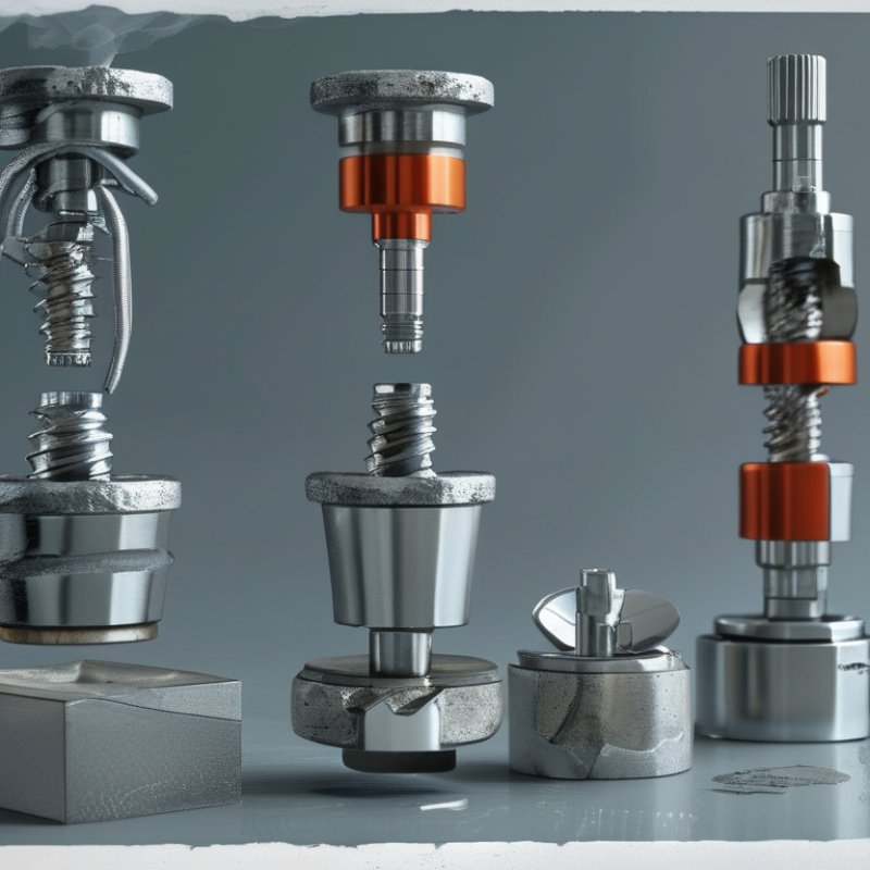
Top Reasons Why Orthopedic Implants Sometimes Fail

What Orthopedic Implant Failure Means and How It Is Identified

Orthopedic implant failure does not always mean the device has broken. It may mean the implant no longer performs safely or effectively. A loose joint component can cause deep pain, clicking, or reduced movement. Infection may appear as warmth, drainage, swelling, or persistent fever. Sometimes, symptoms are surprisingly mild.

Clinicians identify failure through a careful history and physical examination. They ask when pain began, whether it worsens during activity, and how function has changed. X-rays may show migration, bone loss, wear, or a fracture around the implant. CT scans can clarify complex bone changes. Blood tests may support an infection assessment, but normal results do not always exclude one. Joint-fluid analysis may be needed when infection remains uncertain.

Failure can develop from repeated stress, poor bone integration, trauma, infection, or gradual material wear. Patient factors also matter, including bone quality, smoking, weight, and activity level. Still, assigning blame too quickly is a mistake. Pain can come from muscles, nerves, or nearby joints instead. A second clinical opinion may reveal overlooked details. Comparing current images with earlier scans often provides the clearest evidence. Sudden pain after a fall, new deformity, fever, or inability to bear weight requires prompt medical evaluation.

How Implant Design, Materials, and Placement Affect Longevity

Top Reasons Why Orthopedic Implants Sometimes Fail

How Implant Design, Materials, and Placement Affect Longevity

An orthopedic implant can fail when its design does not match the patient’s anatomy, activity level, or bone quality. A joint component may carry repeated loads during walking, climbing, or lifting. Small design differences can change how stress moves through surrounding bone. Poor load distribution may cause loosening, bone loss, or persistent pain over time.

Material selection also affects durability. Metals, ceramics, and medical-grade polymers each respond differently to friction, impact, moisture, and body chemistry. Surface wear can release tiny particles, which may trigger inflammation around the implant. Corrosion is less common with modern materials, but it remains a concern when incompatible components contact each other. No material is perfect. Long-term performance depends on the entire implant system, not one attractive specification.

Placement is equally important. Even a well-designed implant can perform poorly if alignment, depth, or fixation is inaccurate. Surgeons use imaging, surgical guides, and careful soft-tissue balancing to improve positioning. However, anatomy varies, and real operations rarely match textbook conditions exactly. Early movement, infection, falls, smoking, obesity, and delayed bone healing can further reduce stability. Follow-up appointments help detect changes through physical examinations and imaging. In practice, implant longevity reflects both engineering and biology. Sometimes, clinicians must reassess an original decision when symptoms persist without a clear explanation.

The Main Biological and Mechanical Causes of Implant Failure

Top Reasons Why Orthopedic Implants Sometimes Fail

The Main Biological and Mechanical Causes of Implant Failure

Orthopedic implants can fail because the body and the device interact in complex ways. Infection is one serious biological cause. Bacteria may enter during surgery or spread through the bloodstream later. Even a small infection can cause pain, swelling, drainage, or loosening. Blood tests, imaging, and fluid analysis help clinicians investigate these warning signs.

Bone quality also matters. Weak bone may not provide enough support for stable fixation. Some patients develop an inflammatory reaction to wear particles, which can gradually damage nearby bone. Healing may also slow because of smoking, diabetes, poor nutrition, or certain medications. These risks deserve honest discussion before surgery.

Mechanical problems often develop through repeated stress. Excessive loading, poor alignment, falls, or incomplete bone attachment can create abnormal forces. Over time, parts may loosen, fracture, or wear unevenly. A patient may notice new pain during stairs, a change in walking, or unusual clicking. In clinical practice, symptoms and imaging do not always agree. A clean X-ray does not always mean the implant is functioning normally. We can miss early problems. Careful follow-up, comparison with earlier images, and an experienced orthopedic assessment remain essential. Regular review can identify small changes before they become major failures.

How Patient Health and Daily Activities Influence Implant Performance

Orthopedic implants can fail when patient health and everyday loading work against healing. The National Joint Registry’s 21st Annual Report shows that many common hip and knee implants have roughly 3–5% revision risk within ten years. That risk is not identical for every patient.

Body weight matters. The American Academy of Orthopaedic Surgeons reports that obesity increases infection, blood-clot, and revision risks after joint replacement. Excess weight also increases force across the implant during stairs, squats, and rising from a chair.

Diabetes can slow wound healing, while smoking reduces blood flow and may weaken bone integration. Small habits matter.

Daily behavior can quietly change implant performance. High-impact running, repeated jumping, and unsafe lifting may overload surrounding bone or cause falls. Yet complete inactivity is not harmless. Weak muscles can make walking unstable and increase joint stress. The AJRR Annual Report, which tracks millions of hip and knee procedures, emphasizes long-term monitoring because implant outcomes vary by patient factors and activity levels. Patients should follow rehabilitation plans, maintain a healthy weight, control blood sugar, and report persistent swelling, warmth, or new pain. Recovery is not perfectly linear. Some advice may need adjustment, and patients should question routines that feel increasingly difficult.

How Doctors Evaluate, Treat, and Prevent Failed Orthopedic Implants

Top Reasons Why Orthopedic Implants Sometimes Fail

When an orthopedic implant causes pain, doctors first examine the patient’s movement, swelling, skin temperature, and surgical scar. They also ask when the pain began and whether it worsens during walking or rest. A standing X-ray may reveal loosening, bone loss, misalignment, or a fracture near the implant. Sometimes, CT or MRI provides clearer details. No single test is perfect. That uncertainty matters.

Doctors may order blood tests for inflammation and infection. In selected cases, they withdraw joint fluid with a needle for laboratory testing and bacterial cultures. A normal result does not always exclude a low-grade infection. Careful review of earlier scans, operative notes, medications, and medical conditions can reveal overlooked risks. The evaluation should fit the individual, not follow a rigid checklist.

Treatment depends on the cause and the patient’s overall health. Infection may require antibiotics, surgical cleaning, or staged implant replacement. Loosening or severe wear may require revision surgery, bone grafting, and longer rehabilitation. A small fracture can sometimes receive protection and close monitoring, but unstable fractures often need fixation. Prevention begins before surgery through accurate planning, appropriate implant positioning, infection-control measures, and attention to bone quality. Patients can support recovery by following weight-bearing instructions, managing diabetes, avoiding tobacco, and attending follow-up visits. Even experienced teams can miss subtle warning signs, so persistent pain deserves a fresh assessment rather than reassurance alone.

Top Reasons Why Orthopedic Implants Sometimes Fail - How Doctors Evaluate, Treat, and Prevent Failed Orthopedic Implants

Failure Mode or Risk Factor Typical Clinical Features How Doctors Evaluate It Common Treatment Approach Prevention and Risk Reduction Urgency
Aseptic loosening Progressive activity-related pain, reduced function, or new pain after an initially successful recovery. The implant may lose stable fixation without infection. History and physical examination; comparison of serial X-rays for radiolucent lines, migration, subsidence, or changes in alignment. CT or other imaging may be used when plain radiographs are inconclusive. Infection is assessed before revision. Observation may be appropriate for minor, stable findings. Symptomatic loosening or implant migration commonly requires revision surgery, removal of loose components, and restoration of bone support when needed. Accurate implant positioning, appropriate fixation technique, protection of bone quality, weight management, smoking cessation, and follow-up when pain or function worsens. Usually planned
Periprosthetic joint infection Persistent wound drainage, increasing pain, redness, warmth, swelling, fever, drainage, or unexplained stiffness. Some infections are low-grade and may present only with chronic pain or loosening. Examination, blood tests such as C-reactive protein and erythrocyte sedimentation rate, and joint aspiration for cell count, differential, and culture when indicated. Imaging helps assess the implant and surrounding bone. Early infections may be treated with surgical irrigation and debridement plus targeted antibiotics when the implant is stable. Chronic or established infections may require one- or two-stage revision, removal of components, and prolonged antimicrobial treatment. Preoperative screening and treatment of active infections, optimized diabetes control, smoking cessation, appropriate skin preparation and perioperative antibiotics, and careful wound monitoring. Prompt assessment
Periprosthetic fracture A fracture around or near the implant, often associated with a fall, trauma, osteoporosis, or weakened bone. Symptoms commonly include sudden pain, swelling, bruising, deformity, and inability to bear weight. Urgent physical examination and X-rays in multiple views. CT may define fracture extension, bone loss, and implant stability. The stability of the implant and the quality of the remaining bone guide treatment. Stable, nondisplaced fractures may sometimes be managed with protected weight-bearing and close imaging. Displaced fractures, unstable implants, or poor bone support often require fixation, revision components, or both. Fall prevention, osteoporosis assessment and treatment, strength and balance training, appropriate activity progression, and management of modifiable risks such as smoking. Urgent assessment
Dislocation or instability Sudden pain, a visible or palpable change in joint position, inability to move normally, limb shortening, or repeated episodes of giving way. It is most commonly discussed after joint replacement. Physical examination and urgent X-rays to confirm alignment. CT may identify component position, bone defects, or fractures. Doctors also review soft-tissue tension, implant design, activity, and previous operations. Initial treatment may include a closed reduction and temporary precautions. Recurrent instability may require bracing, correction of component position, revision of selected components, or soft-tissue reconstruction. Accurate component orientation, restoration of soft-tissue tension and limb length, adherence to early movement precautions, muscle strengthening, and individualized risk assessment. Urgent assessment
Wear, debris, and osteolysis Gradual pain, swelling, reduced motion, or implant loosening caused by wear particles and bone loss around the implant. Symptoms may be mild despite progressive radiographic changes. Serial X-rays assess alignment, wear, and bone loss. CT or metal-sensitive MRI may be useful for complex anatomy, soft-tissue reactions, or hidden osteolysis. Infection and other causes of pain must be excluded. Small, stable, asymptomatic findings may be monitored. Progressive bone loss, symptomatic wear, or instability may require revision of worn components and treatment of the bone defect. Appropriate implant selection and positioning, avoidance of excessive impact, regular follow-up for high-risk patients, and early investigation of new symptoms. Depends on progression
Malposition or incorrect alignment Persistent pain, abnormal gait, limited motion, instability, impingement, leg-length difference, or uneven loading after fixation or joint replacement. Clinical assessment of alignment, range of motion, gait, and limb length. Weight-bearing X-rays and, when necessary, CT are used to assess component orientation, fracture reduction, and mechanical alignment. Minor symptoms may be treated with rehabilitation and observation. Mechanical problems causing pain, instability, poor function, or accelerated wear may require corrective or revision surgery. Preoperative planning, patient-specific anatomy assessment, intraoperative alignment checks, appropriate component sizing, and postoperative imaging when clinically indicated. Timely assessment
Nonunion or delayed union after fracture fixation Persistent pain at the fracture site, lack of expected functional improvement, abnormal motion, or failure of the fracture to show progressive healing on imaging. Serial X-rays and CT to evaluate bridging bone, fracture gaps, alignment, and implant integrity. Doctors review smoking, nutrition, medications, infection, blood supply, and medical conditions that impair healing. Treatment may include improved immobilization, bone-stimulation strategies in selected cases, correction of metabolic factors, revision fixation, bone grafting, and treatment of infection if present. Stable fixation, preservation of blood supply, appropriate weight-bearing instructions, adequate protein and vitamin intake, smoking cessation, and management of diabetes or osteoporosis. Timely assessment
Implant or fixation failure Breakage, bending, migration, screw loosening, or loss of fixation. It may result from excessive mechanical load, poor bone quality, nonunion, incorrect alignment, or repeated stress over time. Physical examination and comparison of serial radiographs to identify broken hardware, loss of reduction, migration, or changes in alignment. CT and laboratory testing may be needed to assess bone healing and exclude infection. Stable failures may occasionally be observed under specialist supervision. Mechanical instability, significant pain, deformity, or nonunion often requires revision fixation and management of the underlying cause. Appropriate fixation strategy, accurate reduction and alignment, protection of the repair during healing, treatment of osteoporosis, smoking cessation, and scheduled follow-up imaging. Depends on stability
Soft-tissue irritation or impingement Localized pain, tenderness, snapping, bursitis, tendon irritation, or discomfort during specific movements. Symptoms may occur even when the implant is mechanically stable. Physical examination, activity-related assessment, and targeted imaging. X-rays evaluate implant position; ultrasound or MRI may assess tendons, fluid collections, and other soft tissues. Activity modification, physical therapy, anti-inflammatory treatment when medically appropriate, and treatment of associated bursitis or tendon disorders. Persistent mechanical impingement may require surgical correction. Correct implant placement, appropriate component sizing, progressive rehabilitation, avoidance of premature overload, and evaluation of persistent focal pain. Usually nonurgent
Patient-related biological or mechanical factors Risk of failure increases with poor bone quality, obesity, smoking, uncontrolled diabetes, immune suppression, high-impact loading, poor adherence to restrictions, or untreated dental and skin infections in selected situations. Doctors review medical history, medications, nutritional status, bone-health indicators, glycemic control, activity demands, wound status, and rehabilitation progress. Management focuses on correcting modifiable risks, treating underlying disease, adjusting rehabilitation, and selecting observation or revision treatment according to the specific implant problem. Shared decision-making, preoperative optimization, smoking cessation, diabetes and osteoporosis management, infection prevention, realistic activity goals, and long-term follow-up. Long-term prevention
Clinical note: New or worsening pain, fever, wound drainage, sudden deformity, loss of function, or inability to bear weight should be assessed promptly by an orthopedic professional. Implant failure cannot be confirmed from symptoms alone; diagnosis generally requires clinical examination, imaging, and laboratory or joint-fluid testing when infection is possible.

FAQS

: What does orthopedic implant failure mean?

: It does not always mean the implant has broken. It may no longer work safely or effectively. Loosening, wear, infection, bone loss, or nearby fractures can cause failure.

What symptoms may suggest an implant problem?

Deep pain, clicking, reduced movement, swelling, warmth, or drainage may appear. Some problems cause surprisingly mild symptoms. New pain during stairs deserves attention.

How do clinicians identify implant failure?

They review your symptoms, activity changes, and medical history. A physical examination checks movement, stability, swelling, and tenderness. X-rays may show migration, wear, bone loss, or fractures.

Can normal blood tests rule out infection?

No. Normal results do not always exclude infection. Clinicians may combine blood tests, imaging, and joint-fluid analysis. Persistent fever, warmth, or drainage needs prompt evaluation.

Why can an implant become loose?

Repeated stress, poor bone attachment, infection, trauma, or material wear may contribute. Weak bone can provide limited support. Inflammatory reactions may also damage nearby bone over time.

How do health conditions affect implant performance?

Smoking can reduce blood flow and weaken bone integration. Diabetes may slow wound healing. Poor nutrition, excess weight, and some medicines can increase risks.

Can daily activities damage an orthopedic implant?

Repeated jumping, high-impact running, unsafe lifting, or falls may overload surrounding bone. However, complete inactivity can weaken muscles. A balanced rehabilitation plan matters.

Does pain always mean the implant has failed?

No. Pain may come from muscles, nerves, or nearby joints. Symptoms and X-rays do not always agree. We can miss early problems.

When should someone seek urgent medical care?

Seek prompt care after sudden pain, a new deformity, fever, or inability to bear weight. Do not wait for symptoms to become severe. A second opinion may reveal overlooked details.

How can patients monitor an implant over time?

Attend follow-up visits and compare new images with earlier scans. Report persistent swelling, warmth, clicking, or worsening movement. Recovery is not perfectly linear. Some routines may need adjustment.

Conclusion

Orthopedic implant failure occurs when an artificial joint, bone fixation device, or other implant no longer performs as intended. Why do orthopedic implants sometimes fail? The causes may include poor alignment, inadequate fixation, material wear, mechanical loosening, infection, bone loss, or the body’s unfavorable biological response. Implant design, material strength, surface features, and surgical placement all influence how well an implant withstands pressure and movement over time.

Patient health and daily habits also affect implant performance. Conditions such as weakened bones, diabetes, excess weight, smoking, or poor nutrition may slow healing and increase complications, while high-impact activities can place excessive stress on the device. Doctors identify failure through symptoms, physical examinations, imaging tests, and sometimes laboratory analysis. Treatment depends on the cause and may involve observation, medication, revision surgery, infection control, or implant replacement. Careful planning, proper surgical technique, follow-up visits, healthy lifestyle choices, and realistic activity limits can help reduce the risk of failure and improve long-term outcomes.

Aria

Aria

Aria is a dedicated marketing professional with a deep passion for innovative strategies and a keen understanding of our company's product offerings. With a wealth of experience in the industry, Aria excels at crafting engaging content that highlights the unique features and benefits of our......