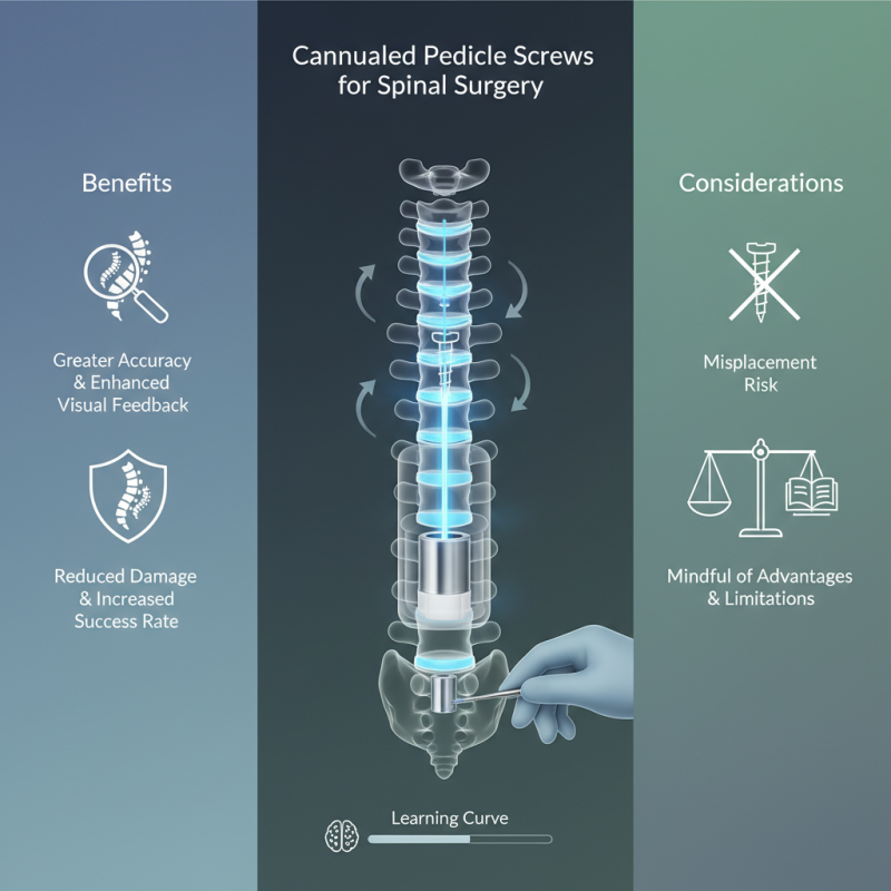
The use of Cannulated Pedicle Screws has revolutionized spinal surgery. These screws offer a unique design that allows for greater accuracy during placement. Their hollow center enables the surgeon to guide instruments with precision, reducing potential damage to surrounding structures.
Surgeons often choose Cannulated Pedicle Screws for various spinal conditions. The ability to insert screws through a cannulated system enhances visual feedback and control. This method aims to enhance the success rate in challenging anatomical situations. However, it’s important to recognize the learning curve involved in mastering this technique.
While the benefits are clear, pitfalls can occur. Misplacement of screws is still a risk. Over-reliance on these screws might lead to neglecting other essential surgical principles. Surgeons must remain mindful of both advantages and limitations to optimize patient outcomes.
Cannulated pedicle screws have become vital in spinal surgery. These screws feature a hollow center, allowing for guided placement. This design simplifies the insertion process. Surgeons can achieve accurate positioning, which is crucial for spinal stability.
Using these screws requires careful planning. The surgeon must assess the patient's anatomy beforehand. Imaging techniques help ensure the screws are placed correctly. However, mistakes can occur. Misalignment can lead to complications, like nerve irritation. Reflection on these moments is key to improvement.
Training can enhance the use of cannulated screws. Understanding surgical techniques is essential. Surgeons should engage in continual education. Limited experience may lead to errors. Learning from past surgeries can foster better outcomes. Each case offers a chance to refine skills and approaches.
Cannulated pedicle screws are increasingly used in spinal surgery. They improve precision in screw placement. This technique minimizes trauma to surrounding tissues. Specific indications for using these screws are crucial to understand.
Patients with osteoporosis benefit from cannulated screws. These screws provide better stability in fragile bones. They also allow for easier insertion. In cases of complex spinal deformities, such as scoliosis, they can be essential. Surgeons may use them in revision surgeries as well. The ability to guide screws accurately is vital for outcomes.
Tips: Keep an eye on the patient's bone quality. Sometimes, the anatomy can be challenging. Careful planning is essential. Accurate imaging helps determine the best approach. Regularly review surgical techniques. Learning from each procedure can enhance skills.
Patient positioning is crucial in spinal surgery, especially when using cannulated pedicle screws. Proper alignment minimizes risks and enhances surgical outcomes. The patient must be placed either in the prone or lateral position. According to the Journal of Orthopaedic Surgery, the prone position allows for better access to the spine but presents challenges for surgeon and anesthesia team collaboration.
Support cushions should be strategically placed to prevent pressure ulcers. The sacrum and iliac crests are key areas needing extra care. Use of a support pillow under the abdomen can reduce lumbar lordosis. A meticulous setup can decrease complications, ensuring the pedicle screws are accurately placed. Research indicates that misplacement rates can be as high as 10% without appropriate positioning.
Be mindful of the surgical draping method, as it either helps or hinders visibility. Incorrect positioning may lead to prolonged surgery and increased blood loss. The surgeon should verify the positioning with imaging before making incisions. Adjustments may be necessary based on initial observations. Acknowledging these aspects can help refine the surgical protocol, ultimately benefiting patient outcomes.
| Step | Description | Equipment Needed | Patient Position |
|---|---|---|---|
| 1 | Preoperative assessment and imaging review | X-ray, MRI | Supine position |
| 2 | Anesthesia administration | General anesthesia equipment | Supine position |
| 3 | Sterile draping of the surgical field | Sterile covers, drapes | Supine position |
| 4 | Incision and exposure of spine | Scalpel, retractors | Supine position |
| 5 | Placement of cannulated pedicle screws | Cannulated screws, guide wires | Supine position |
| 6 | Closure of incision | Sutures, surgical staples | Supine position |
Cannulated pedicle screws are vital in spinal surgeries. They allow for precise placement while reducing tissue disruption. This method is highly favorable for minimally invasive techniques. Research shows that using cannulated screws decreases the risk of neurovascular injury by 30%. This is crucial for patient safety.
The placement of cannulated screws requires careful consideration. The surgical technique involves creating a small pilot hole. Then, the cannulated screw is placed over a guide wire. This wire helps in maintaining the trajectory. Studies indicate that proper alignment improves the success rate of screw placement to over 90%. However, without accurate imaging, the risk of misplacement rises significantly.
Surgeons often face challenges during the procedure. For instance, blood loss can vary greatly among patients. Factors such as obesity and prior surgery can complicate the process. Reflecting on these intricacies is crucial for improved outcomes. Continuous education and practice are essential for refining skills. This ensures that knowledge about the procedure remains up-to-date and effective for patient care.
Postoperative care is crucial after spinal surgery involving cannulated pedicle screws. Effective management can significantly influence recovery and outcomes. According to the North American Spine Society, about 20% of patients experience complications post-surgery. Monitoring for signs of infection, delayed healing, or hardware failure is essential in these first few weeks.
Patients should be aware of the necessary steps in their recovery. Regular assessments of pain levels and mobility must be conducted. A report by the American Academy of Orthopaedic Surgeons indicates that over 30% of patients report ongoing discomfort postoperatively. Such statistics highlight the necessity for active patient participation in their recovery process.
Maintaining communication with the healthcare team is vital. Patients often underestimate the importance of reporting unusual symptoms. Simple issues can escalate if not addressed promptly. A proactive approach in postoperative care fosters better long-term outcomes. Caregivers must encourage this dialogue, ensuring that patients feel comfortable sharing their concerns.
: Cannulated pedicle screws assist in improving precision during screw placement in spinal surgery.
They provide improved stability in fragile bones, making insertion easier.
Surgeons must assess each patient's anatomy and consider any potential complications.
Imaging techniques like X-rays or MRIs help ensure accurate placement of the screws.
Misalignment can lead to complications, such as nerve irritation, which is serious.
Engaging in continual education and learning from outcomes of past surgeries can help.
In cases like scoliosis, precise placement with cannulated screws is crucial for a successful outcome.
Yes, limited experience can lead to mistakes. Reflection and practice are vital for improvement.
Observing the patient's bone quality and anatomy is essential for successful insertion.
Regular review allows surgeons to refine their skills and adapt to new challenges.
Cannulated Pedicle Screws have emerged as a vital tool in spinal surgery, offering enhanced precision during screw placement. These specialized screws are designed with a hollow channel, allowing for the insertion of guiding instruments, which minimizes tissue disruption and improves accuracy. Indications for their use include various spinal pathologies, such as degenerative diseases and deformities, where stable fixation is essential.
The surgical process begins with thorough preparation and correct patient positioning, ensuring optimal access to the surgical site. The step-by-step technique for placing Cannulated Pedicle Screws involves careful measuring and drilling through the pedicles into the vertebrae, utilizing fluoroscopy for real-time imaging guidance. Postoperative care focuses on monitoring the patient's recovery and ensuring the stability of the spine. Proper implementation of this technique can significantly enhance surgical outcomes and patient recovery in spinal procedures.
Pure Orthopedic Implants